
A colored image of a pea aphid embryo taken at the University of Miami Imaging Core Facility received honorable mention in the 2014 Nikon Small World Photomicrography Competition. The event showcases “the beauty and complexity of life as seen through the light microscope.”
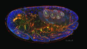 |
| Image of a pea aphid embryo taken at the University of Mami Imaging Core Facility, Department of Biology. Credit: Hsiao-Ling Lu |
This year is the 40th anniversary of the competition. The photomicrography competition received more than 1,200 entries from 79 different countries. Only 12 received honorable mention. The winners were announced on Thursday, October 30, 2014.
The image was taken by Hsiao-Ling Lu, a doctoral student at the National Taiwan University. Lu took the image in 2013 at the University of Miami (UM), when she was a visiting fellow from June 2013 to June 2014, in the Department of Biology in the College of Arts and Sciences.
“I feel very honored and excited to receive this distinction,” Lu said. “I adore the images that exhibit the beauty of the cell, the organ or the embryo,” she added. “I hope my images can provide biological information at different levels of comprehension and appreciation.”
Lu is a “highly skilled aphid developmental biologist,” according to Alex Wilson, an associate professor of biology in the UM College of Arts and Sciences and director of the UM lab where Lu worked.
“Her visit resulted in an amazing expansion of my group’s research on the evolution of symbiotic interactions by facilitating novel investigations of host-symbiont developmental integration,” Wilson said.
The images were taken using confocal microscopy. This technique allows users to produce optical sections of specimens and reconstruct three-dimensional structures from the obtained images, without cutting the sample. It allows researchers to precisely locate proteins and structures in living and fixed specimens.
“The Imaging Core Facility serves a broad community of faculty and students from many disciplines, including biology, geology, marine sciences, chemistry and medicine,” said James Baker, research assistant professor and manager of the Imaging Core Facility at UM Department of Biology. “It has led to a number of interdisciplinary publications and we hope to expand those collaborations in the future.”
October 30, 2014
