Although he’s only in his third year of undergraduate studies at the University of Miami, Sumedh Shah, a biology major in the College of Arts and Sciences, is already conducting groundbreaking research on glioblastoma, an aggressive form of brain cancer that killed his father.
Shah uses a powerful electron microscope – capable of magnifying objects up to 1.2 million times – to view the cancer cells up close, and see how their structures differ from normal cells.
“The electron microscope allows us to cut a single layer of cells into segments just 60 nanometers thick,” he said. A nanometer is one-billionth of a meter. Shaw added that the technique requires extreme precision, eye-hand coordination and time.
Shah is honing these skills in the Techniques in Electron Microscopy course, taught by Jeffrey Prince, an associate professor of biology in the College of Arts and Sciences.
Prince’s class is one of the most exclusive on campus. More than 40 students applied for just seven spots. Prince personally interviews each student, and acceptance is based upon their grades, courses taken and additional factors.
“This class is a reward for hard-working students,” he said, adding that the electron microscope is worth close to $1 million, and the lab work involves toxic, even explosive, chemicals. “They have to be responsible and willing to put in the time and effort. The intent of this class and laboratory from the beginning has been to provide UM students with a trait that allows them to be heads above all other applicants for a professional position.”
Prince and his students invited UM President Donna E. Shalala and Vice President for Student Affairs Patricia Whitely to the lab on November 17, to learn about their research and how the electron microscope is expanding the horizons of science.
Prince said, “This class is not a show and tell; it is hands-on. And for the last 30 years, students have stepped up and done it – with excellence.”
Shah – who serves as Prince’s teaching assistant – is enrolled in the Honors Program in Medicine (HPME), through which he will earn a B.S. in biology from the College of Arts and Sciences and an M.D. from the Miller School of Medicine in seven years.
He noted that all of Florida’s research institutions have electron microscopes on campus, but UM is the only institution that allows undergraduate students to use the equipment. “We are learning a skill that most do not get until graduate school,” he said.
Two more of Prince’s students are also working on innovative collaborative research projects.
Senior Neville Patel is investigating Charcot-Marie-Tooth (CMT) disease, an inherited neurological disorder that affects about 1 in 2,500 people in America. Involving both motor and sensory nerves, CMT causes weakness in the foot and lower leg muscles. Patel examines the affects of genes that cause CMT with the electron microscope. He is applying to medical schools for fall of 2015, and is an author on a paper that has been submitted to the journal Nature Genetics.
Senior Mateuzs Graca has been working with the electron microscope since he was a first-year student. He is studying retinitis pigmentosa, an inherited degenerative eye disease that causes blindness. Researchers at UM’s Bascom Palmer Eye Institute identified the gene that causes retinitis pigmentosa in 2011; Graca is examining those cells with the electron microscope, seeking a cure.
Graca is currently interviewing for medical school for fall of 2015. He said, “Just days ago, an interviewer asked about my background as an applicant. I told her about this research. The interviewer was very impressed by this experience.”
Other current students are: pre-med sophomore Natalie Flores, who will be taking over Graca’s research when he graduates; junior Elizabeth Guirado, who plans to earn a joint D.M.D./Ph.D. in dentistry; junior Eric Keen, who received honorable mention for a 2014 Goldwater Scholarship; first-year student Kasey Markel, who said that the electron microscopy lab was a major factor in his decision to attend UM and will be helping Shah with his research next semester; junior Katelyn O’Neill, who will also be working on retinitis pigmentosa research; senior Dominika Swieboda, who is planning to pursue a Ph.D. in microbiology; and sophomore Mason Schecter, a third generation ’Cane majoring in biology, physics and chemistry.
Keen said, “Our work is complementary with what is going on at the medical school. What is unique is that we can look at cells directly. This is a piece of the puzzle that is very relevant today.”
He is interested in virology, and will be using the electron microscope for his ongoing research on viruses that attack bacteria. He said, “Once you show that you are able to use the technology effectively and safely, Professor Prince gives you the freedom to pursue research that is meaningful to you.”
Shalala congratulated the students on their work, adding, “I think you are all privileged to be able to participate in such a class.”
Shah concurred. “This class has made my time at UM worth it,” he said.
Electron microscopes are costly to procure, and expensive to maintain. The annual service contract for the scopes and other gear in the Dauer Lab is $60,000. This ensures that a technician will arrive within one to two days when problems occur with the delicate apparatus.
The grant funding the Dauer Lab service contract ends in March 2015, and the Department of Biology is unable to absorb the maintenance costs if other resources cannot be identified. The student research projects – and the Dauer Electron Microscopy Lab itself – risk closure. Prince said that 70 percent of the nation’s electron microscopes have been shut down due to lack of funding for service contracts.
“The electron microscope must be consistently available for effective teaching and research,” he said. “The initial images produced by the first-year students and the research conducted by the project students are remarkable.”
Melissa Peerless can be reached at 305-284-2485.
Science and Technology
Lab Provides Unique Opportunity
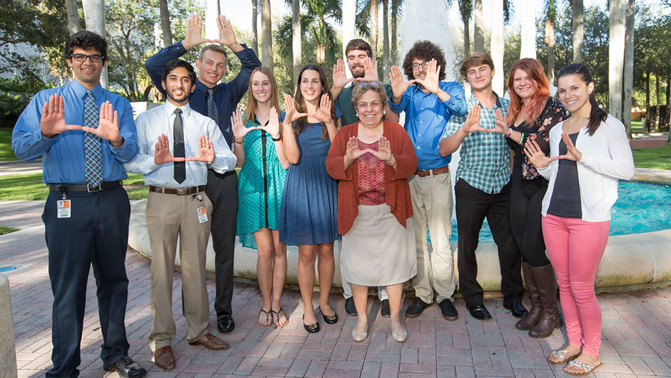
Students in the Techniques in Electron Microscopy course with UM President Donna E. Shalala.
University of Miami
Coral Gables, FL 33124
305-284-2211